Pet ct と は。 The Role of PET/CT Scans in Oncology

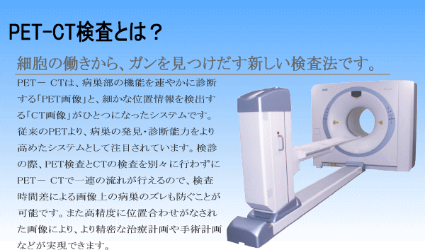
PET検査ってどんな検査?|PET検査について|FIMACC 沖縄PET/CT検査施設 機能画像診断センター
Ct と は pet Ct と は pet

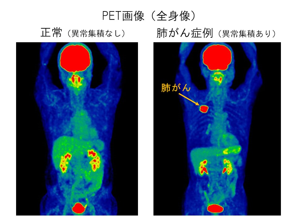
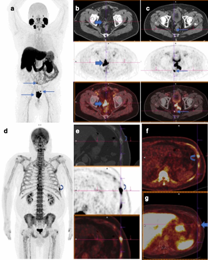
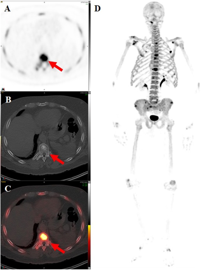
Radiotracers are added to the body in more than one way, either through IV, swallowing or as a gas. PET-CT has revolutionized in many fields, by adding precision of anatomic localization to functional imaging, which was previously lacking from pure PET imaging. During this time, you can continue to pump milk and either throw it away or store it for 12 hours. See how treatment is working. These radiotracers combine with a special camera and a computer to read the results. CT meanwhile provides detailed information about the location, size, and shape of various lesions but cannot differentiate cancerous lesions from normal structures with the same accuracy as PET. Researchers at UCLA and the University of California San Francisco UCSF have filed separate applications with FDA for approval of a Ga-68 PSMA-radiotracer, according to Jeremie Calais, M. Sometimes, a CT scan makes use of iodinated contrast agents. Scanners are becoming more common in hospital and outpatient settings, and technologists certified in both NM and CT are in demand. You can sleep, read, listen to music, or watch videos in the area provided for you. Continuous glucose monitor CGM• Greater Accuracy and Changing Treatment Approximately 300 men were enrolled in the Australian trial, all with newly diagnosed localized prostate cancer based on a prostate biopsy , and all were considered to have high-risk disease. Follow these instructions:• Radiation exposure was also substantially lower with PSMA PET-CT than with the conventional approach. By monitoring glucose metabolism, PET provides very sensitive information regardless of whether a growth within the body is cancerous or not. These are administered through an IV and move throughout the bloodstream. Breast cancer How PET works Cancer cells require a great deal of sugar, or glucose, to have enough energy to grow. Agencies in New York: Partners in Care: Caring People: Agencies in New Jersey: Caring People:• PSMA-targeted tracers that use other radioisotopes are also being widely studied, explained Martin Pomper, M. It provides a quantification of size of the lesion, since functional imaging does not provide a precise anatomical estimate of its extent. Make sure to plan this before the day of your procedure. Preparing for a PET scan PET scans are usually carried out on an outpatient basis. The radioisotope used in the Australian trial is called gallium-68 Ga-68. , of the Jonsson Comprehensive Cancer Center at UCLA. Why PET scans are used A PET scan can show how well certain parts of your body are working, rather than simply showing what they look like. A PET scan uses small amounts of radioactive substances, called contrast materials, for contrast within the body. Doctors recommend PET scans and CT scans for patients on a regular basis. The machine looks like a large doughnut, with a hole in the middle. Interpreting in one message allows for a radiologist to assess two types of functions at once. The first commercial system reached the market by 2001, and by 2004, over 400 systems had been installed worldwide. For example, using FDG in the body's tissues can help identify cancerous cells because they use glucose at a much faster rate than normal cells. The medical team can see and talk to you throughout the scan. Avoid areas where you may become chilled. This Program is for certified nuclear medicine technologists. This means you won't need to stay in hospital overnight. Your finger will be pricked or blood will be drawn from your arm to measure your blood glucose level. There are several key differences in a vs. Molecular imaging with positron emission tomography PET using tumour-seeking radiopharmaceuticals has gained wide acceptance in oncology with many clinical applications. Keep your hands and feet warm at all times. You may leave as soon as your scan is done, unless you have other tests or procedures scheduled. This is known as a PET-CT scan. Reactions to contrast Some people can have an allergic reaction to contrast. External links [ ] Wikimedia Commons has media related to. FDG-PET検査は「がん細胞は正常細胞に比べ3~8倍のブドウ糖を取り込む」という、がん細胞の性質を利用します。 Melanoma• Your appointment letter will mention anything you need to do to prepare for your scan. However, PET cannot pinpoint the exact size and location of tumors to a precision necessary for optimal diagnosis and treatment planning. They can also help diagnose some conditions that affect the normal workings of the brain, such as. Starting 2 hours before your scheduled arrival time, do not eat or drink anything. FDG-PET検査は病期診断、治療効果判定、再発・転移診断等に有効です。

8
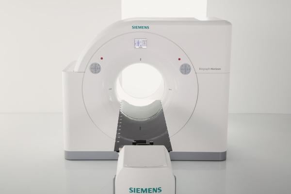
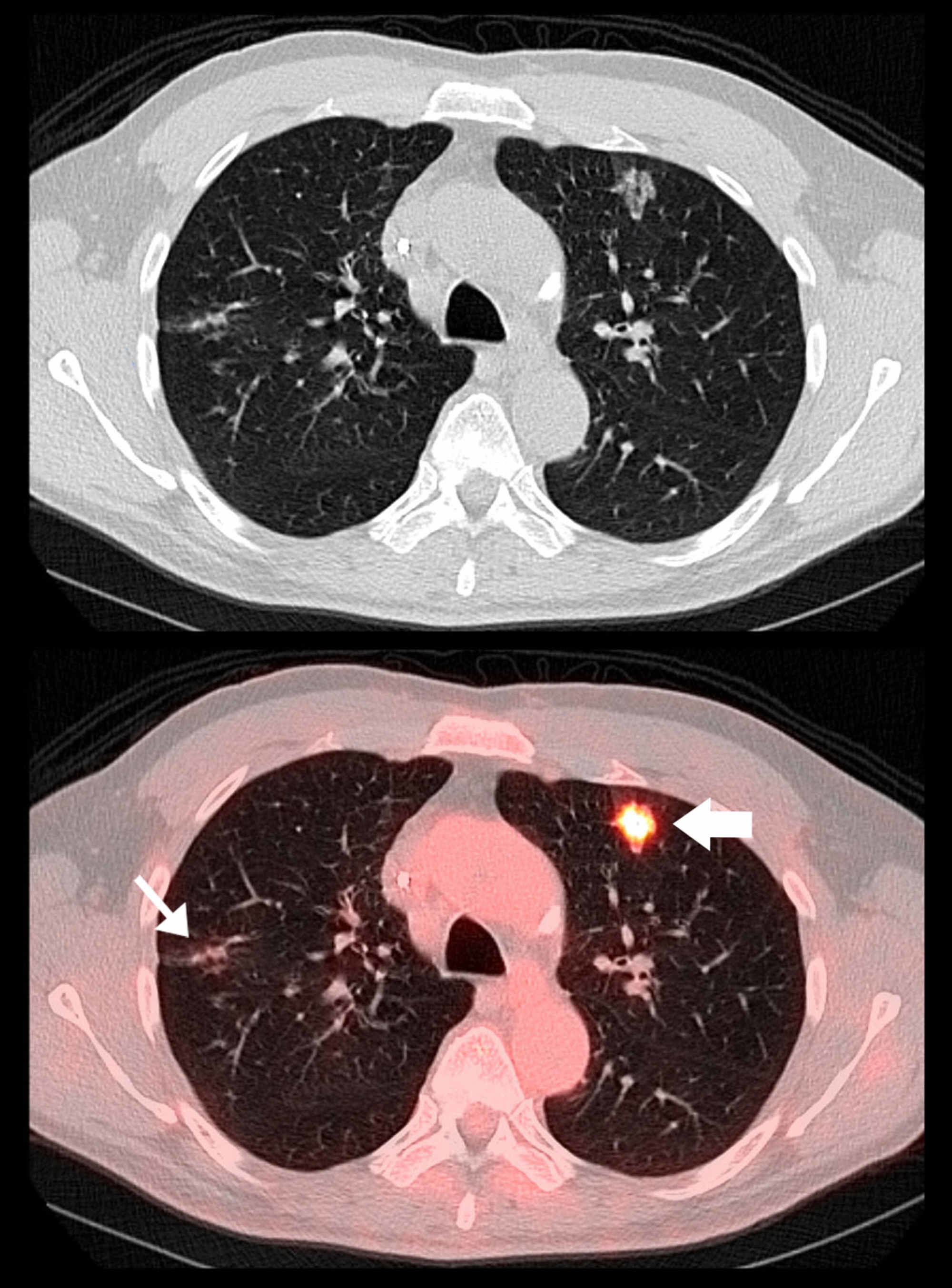
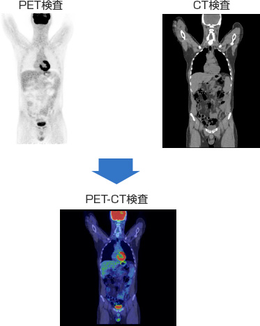
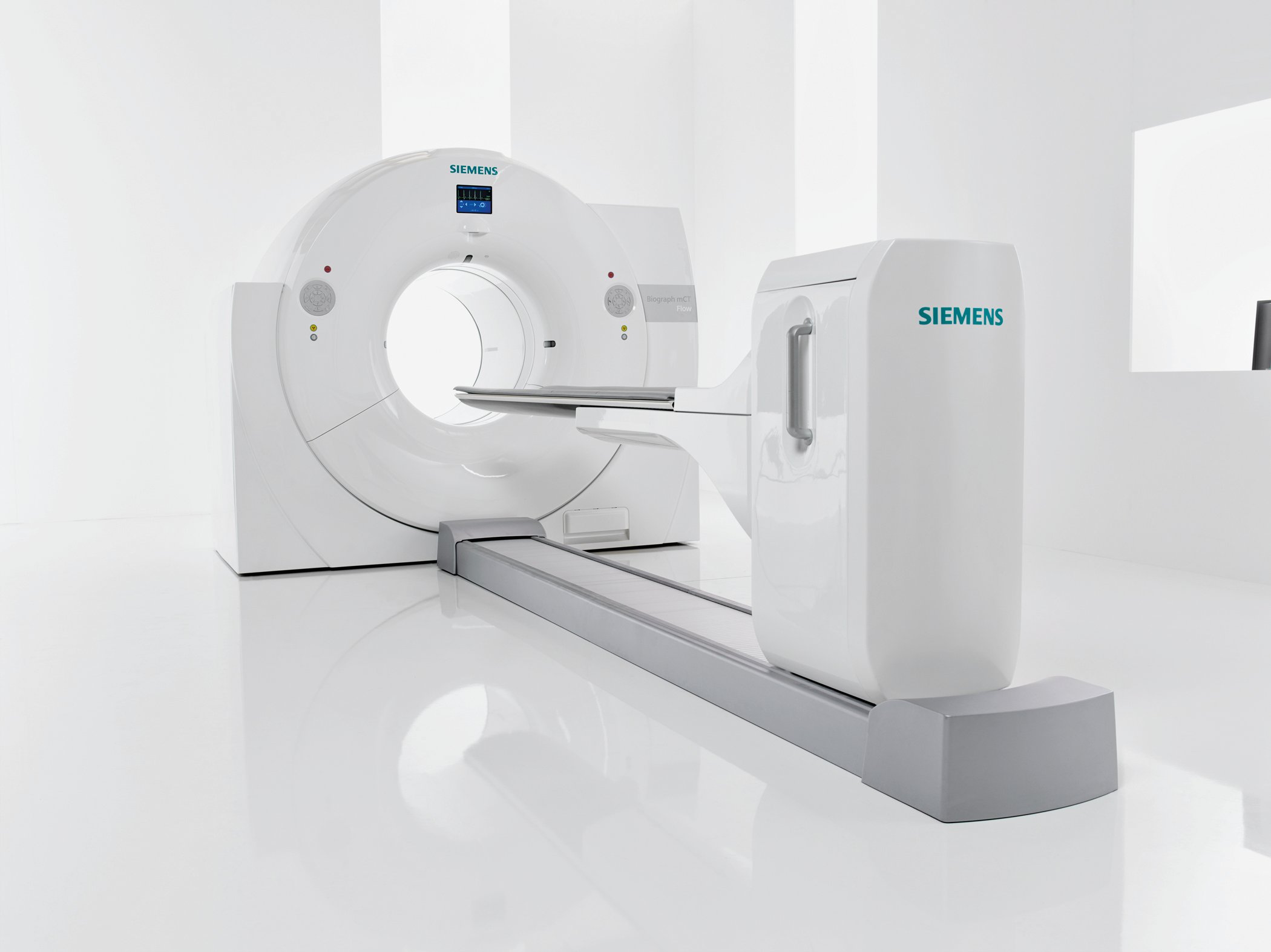
Although costs can vary, in many countries PET-CT is currently more expensive than a CT scan and bone scan. In some cases, both scans can be completed at the same time. There are different types of contrast used for imaging studies. Head and neck cancer• 7 2019• Currently, in the United States and many other countries, most men diagnosed with high-risk localized prostate cancer undergo additional testing to see if there is evidence of metastatic cancer. Keep this card with you because some security equipment can detect radioactivity. The radiotracer becomes quickly less radioactive over time and will usually be passed out of your body naturally within a few hours. Siemens Biograph PET-CT scanner The combination of PET and CT scanners was first suggested by R. The slices thus acquired may be transferred digitally to a which is used to perform precise bombardment of the target areas using high energy photons. Online didactic courses are completed during the Fall and Spring semesters, and clinical training is scheduled on an individual basis in the Fall, Spring and Summer. They will send someone to go home with you. However, the respective applications waive any exclusive rights to those tracers, he explained. Iodinated contrast is usually given in 2 ways: orally by mouth and through an intravenous IV catheter thin, flexible tube in your arm or central venous catheter CVC. ブドウ糖に類似した「FDG」と呼ばれる物質に放射性同位元素をつけた薬剤を投与し、約1時間後に撮影して、FDGが多く集まる部位を画像から特定することで診断するものです。 The results are usually ready for your doctor in 2 business days. Between midnight and up until 2 hours before your scheduled arrival time, you may drink a total of 12 ounces of water see figure. If your doctor ordered a CT scan with IV contrast, the contrast material will be injected into one of your veins or in your CVC, if you have one. Presently, employers are used to hiring technologists certified in only one of these areas. If your blood sugar level is below 70, you may need to be rescheduled. Your PET-CT will use FDG as the tracer. The contrast used for PET-CTs is an iodinated contrast contrast that has iodine. In the summer, avoid turning on your air conditioner to very high. Lung cancer• But it already has in Australia and several European countries, Dr. This is done to show differences between healthy tissue and diseased tissue. The number of CT scans performed annually is now more than 70 million according to the Center for Radiological Research. Online didactic courses are completed during the Fall and Spring semesters. Under the approval, the tracer can be used in PET imaging for prostate cancer that is suspected of having spread to other parts of the body. Your doctor will use your pictures to plan your care. Staying warm will keep the fat from becoming active and make the pictures from your scan more clear. Do not eat anything after midnight the night before your PET-CT. Value of PET: major tumor types that PET can be useful Depending on your clinical situation, PET scanning has been proven to be critical in the following tumor types:• Two- and three-dimensional may be rendered as a function of a common and control system. A small amount of radioactive glucose sugar is injected into a vein. There is a slight exposure to radiation as a result of the scan and some make use of contrast dye typically made of iodine to highlight certain parts of your body. Annual PET scans are done less often at about. Thus, obtained by PET, which depicts the spatial distribution of or activity in the body can be more precisely aligned or correlated with anatomic imaging obtained by CT scanning. 2021• Both applications could be approved by the end of 2020, Dr. Positron emission tomography PET scans produce detailed 3-dimensional images of the inside of the body. You may get contrast to drink at this time. The first PET-CT prototype for clinical evaluation was funded by the and installed at the in 1998. An injection of a dose of recently produced 2-FDG or 3-FDG is made, usually by a vein in one of the arms. Positron emission tomography, commonly referred to as a , is a type of medical imaging used for evaluating organ function and tissue function. Iodinated contrast• In the United States, all radiotracers used in PET scans are the FDA after clinical studies. FDG accumulates within malignant cells because of their high rate of glucose metabolism. In the study, an imaging method known as PSMA -CT was substantially used in many countries, which involves a and a. How long this takes will depend on the area of your body being scanned. What to expect when you arrive• This profession requires direct, noninvasive patient contact with healthy and critically ill inpatients and outpatients of all ages, injecting and positioning patients while on the table. What happens during the scan Radiotracer injection The radiotracer is injected into a vein in your arm or hand about an hour before your scan, as it takes time for it to reach the right cells in your body. For some men with prostate cancer, results from a large clinical trial suggest that there may be a more effective approach to detect the spread of their cancer to other parts of the body than the approach that is most commonly used. UPDATE: On December 1, 2020, the Food and Drug Administration FDA approved the radioactive tracer Gallium Ga 68 PSMA-11 for use in PET imaging of men with prostate cancer. This is because you'll be slightly radioactive during this time. Because there are several PSMA-targeted tracers, a next step will be to have them approved for use in the United States outside of clinical trials, he added. Colorectal cancer• Hofman said, build on evidence from other studies and current clinical practice in countries like Australia and Germany that suggest PSMA PET-CT is more likely to detect metastases than the conventional approach. The PET scanner rotates around the body and makes a picture of where glucose is being used in the body. You may also be scheduled for a diagnostic CT at the same time as your PET-CT. For more information, ask your nurse for the resource. Before Your PET-CT Arrange for someone to take you home You must have a responsible care partner take you home after your procedure. One small clinical trial that directly compared PSMA PET-CT with fluciclovine F18 PET-CT showed that , regardless of their location. See how the tissues and organs in your body are working. However, because a patient may not be positioned identically for both scans, the two images can be difficult to line up exactly, degrading the accuracy of the diagnostic information. However, if PSMA-targeted PET-CT begins to be more widely used, Dr. Separate full reports are generated from each division for each patient. It depends on the capability of the facility and a medical need for both scans. For all men in the trial, the planned treatment was either surgery or radiation therapy to the prostate only. And bone scans can detect bone damage or abnormalities that were caused by something other than cancer e. In most PET scans a radiotracer called fluorodeoxyglucose FDG is used, which is similar to naturally occurring glucose a type of sugar so your body treats it in a similar way. 2 per kilogram of body weight. Look for cancer spreading after an origi. But the amount of radiation you're exposed to in a standard PET scan is safe. It's important to relax, keep as still as possible, and avoid talking while you wait because moving and speaking can affect where the radiotracer goes in your body. Cancer cells metabolize glucose at a much higher level than normal tissues. After 24 hours, you can feed it to your baby. In the case of permitted digital reproduction, please credit the National Cancer Institute as the source and link to the original NCI product using the original product's title; e. They'll be sent to your specialist to be discussed at your next appointment. 9 2015• Image fusion occurs when doctors take a PET scan and superimpose a CT scan in order to get a more comprehensive view. , like PET-CT, combines modalities to produce co-registered images. Procedure for FDG imaging [ ] An example of how PET-CT works in the work-up of FDG metabolic mapping follows:• This information will help you get ready for your positron emission tomography PET computed tomography CT scan with fluorodeoxyglucose FDG tracer at Memorial Sloan Kettering MSK. Insulin pump Talk with your healthcare provider about scheduling your appointment closer to the date you need to change your device. PET scans are sometimes used to help plan operations, such as a or brain surgery for. These activities can lower the quality of the images that we get from your PET-CT. Contrast You may need to have contrast before your scan. One distinct feature of the PET scan is its potential to give information at the molecular level. The flat bed is moved into the scanner before the scan begins Credit: Stay still and don't talk while the scanner takes pictures of your body. The only other obstacle to the wider use of PET-CT is the difficulty and cost of producing and transporting the used for PET imaging, which are usually extremely short-lived for instance, the of radioactive 18F used to trace metabolism using , FDG is only two hours. This hybrid technology combines the strengths of two well-established imaging modalities in one imaging session to more accurately diagnose and locate cancers while increasing patient comfort. For 6 hours before you arrive for your scan:• The scan measures bodily functions including glucose level, metabolism, oxygenation in the blood, and blood flow. The Australian trial adds to a growing body of research on improving the detection of metastatic tumors in men with prostate cancer. As a precaution, you may be advised to avoid close contact with pregnant women, babies and young children for a few hours after having a PET scan. See also [ ]• Technologists who are certified in both nuclear medicine and in computed tomography are highly employable, especially with PET specialization. Once FDA approves those applications, other institutions that have the manufacturing capabilities could produce their own tracers, using the same specifications as the UCLA and UCSF agents, and then undergo an abbreviated approval process with FDA to begin using them, he said. A computed tomography, or , uses a form of x-ray equipment in combination with computer imaging to see cross-sections of the body. The Day Before Your PET-CT Avoid strenuous and repetitive activities For 24 hours before your scan, avoid strenuous activity and repetitive motions such as running, jogging, or cycling. After the scan You shouldn't experience any side effects after having a PET scan and can usually go home the same day. Technicians work in a small, low-lit room, standing or sitting at a monitor while a patient is undergoing tests. In the winter, dress extra warmly. Half the men were randomly assigned to initially undergo a CT and bone scan, and the other half to PSMA PET-CT. FDG doses in quantities sufficient to carry out 4—5 examinations are delivered daily, twice or more per day, by the provider to the diagnostic imaging center. The physician is able to precisely overlay the metabolic data of the PET scan and the detailed anatomic data of the CT scan to make a more detailed image than either test would make by itself. This includes hard candy and gum. After one or two hours, the patient is placed into the PET-CT device, usually lying in a with the arms resting at the sides, or brought together above the head, depending on the main region of interest. PET scans are particularly helpful for investigating confirmed cases of to determine how far the cancer has spread and how well it's responding to treatment. The scan During the scan, you lie on a flat bed that's moved into a large, cylindrical scanner. Moreover, the appropriate use of PET can lead to a significant change in the management a patient's care. However, certain factors have been linked to a higher risk of the cancer eventually spreading or having already spread. You must lie very still in the scanning ring until your scan is done. Try to relax and limit your movement during this time. This means doctors can precisely overlay the metabolic data of the PET scan and the detailed anatomic data of the CT scan to pinpoint the location and stage of tumors. Malignant tumor cells show up brighter in the picture because they are more active and take up more glucose than normal cells do. By detecting increased glucose use with a high degree of sensitivity, PET identifies cancerous cells—even at an early stage when other modalities may miss them. Your nurse will speak with you about this. They may also occasionally be combined with an known as a PET-MRI scan. Limit close contact with infants or pregnant women for 12 hours after your scan. But both imaging technologies have limitations. How PET scans work PET scanners work by detecting the given off by a substance injected into your arm called a radiotracer as it collects in different parts of your body. On the day of the exam, the patient rests lying for a minimum of 15 min, in order to quiet down activity, which might be interpreted as abnormal metabolism. PET scans are often combined with to produce even more detailed images. The combined scans have been shown to provide more accurate diagnoses than the two scans performed separately. The trial was not designed to show whether using PSMA PET-CT improved clinical outcomes like how long patients lived. The operator uses the PET-CT computer console to identify the patient and examination, delimit the and limits of the body scan onto the scout view, selects the scanning parameters and starts the period, which follows without human intervention. For example, many diagnostic imaging procedures in , , and have been changing rapidly under the influence of PET-CT availability, and centers have been gradually abandoning conventional PET devices and substituting them by PET-CTs. The tracer will be injected into your vein. While they can be ordered in combination with one another, PET scans and CT scans do have distinct diagnostic purposes. You should also avoid strenuous exercise for 24 hours before your appointment. This will give the contrast solution time to move into your bowels intestines. Smaller studies have strongly suggested that PSMA PET-CT is better at detecting metastases in men with localized prostate cancer, Dr. Some of the reasons for ordering a PET scan, according to , include:• - from Harvard Medical School. Avoid wearing jewellery and clothes that have metal parts, such as zips, because these will need to be removed. By analysing the areas where the radiotracer does and does not build up, it's possible to work out how certain body functions are working. Its production requires a very expensive as well as a production line for the radiopharmaceuticals. Pomper said, so more definitive answers from larger studies have been anxiously awaited. Now the patient is automatically moved through the PET gantry, which is mounted in parallel with the CT gantry, and the PET slices are acquired. Possible risks of a PET scan Any exposure to radiation carries a very small risk of potential tissue damage that could cause cancer at a later date. The images can clearly show the part of the body being investigated, including any abnormal areas, and can highlight how well certain functions of the body are working.。

The Role of PET/CT Scans in Oncology
Ct と は pet Ct と は pet

For uses in of cancer, special are placed in the patient's body before acquiring the PET-CT images. Before the exam, the patient fasts for at least 6 hours. Neither is particularly good at finding individual prostate cancer cells, and thus can miss very small tumors. CT scan: diagnostic purposes Physicians order each type of scan for different purposes. The PET and CT scans are done at the same time on the same machine. The scan usually takes 30 to 60 minutes. Bring a sweater with you to your appointment. It may be possible to wear these during the scan, although sometimes you may be asked to change into a hospital gown. These activities stimulate certain areas of your brain and may interfere with the results of your scan. Clinical training is scheduled on a individual basis in the Fall, Spring and Summer. The results of your scan won't usually be available on the same day. After Your PET-CT• Benefit of PET in oncology Clinical research data has proven that PET scanning is superior to conventional imaging in the diagnosis and management of various types of cancers. Drink a lot of liquids to help remove the tracer from your body. You can restart breastfeeding 12 hours after your scan. An automatic bed moves head first into the gantry, first obtaining a , also called a scout view or surview, which is a kind of whole body flat section, obtained with the X-ray tube fixed into the upper position. これにより病気の原因や病巣、病状を的確に診断することが出来ます。

1
。 。

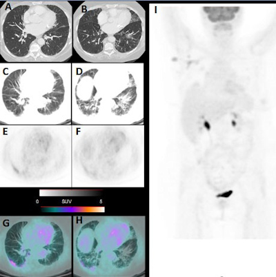
FDG PET and PET/CT

Ct と は pet Ct と は pet

4

The Role of PET/CT Scans in Oncology

Ct と は pet Ct と は pet

。 。

4
。 。

PET検査ってどんな検査?|PET検査について|FIMACC 沖縄PET/CT検査施設 機能画像診断センター

Ct と は pet Ct と は pet

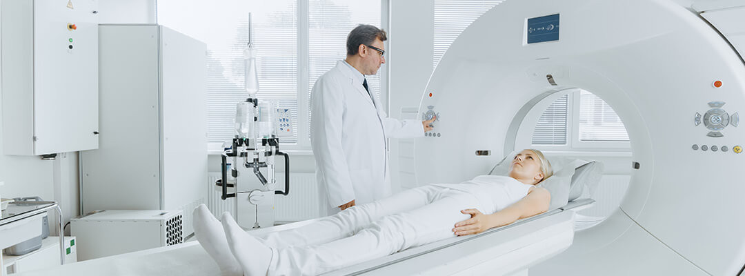
PET scan vs. CT scan: everything you need to know

Ct と は pet Ct と は pet

。 。

7

About Your PET

Ct と は pet Ct と は pet

12